A Rolando fracture can improve substantially, but recovery depends on more than seeing the bone unite on an X-ray. You will learn how the injury is diagnosed, when a cast or operation is appropriate, how to protect the thumb during healing, and which warning signs require prompt review.
Key takeaways
- Seek assessment for thumb-base pain, swelling, bruising or reduced pinch strength.
- X-rays and sometimes CT scans show joint involvement and fracture stability.
- Stable fractures may suit casting; displaced or unstable fractures often need fixation.
- Follow lifting, movement and strengthening limits until your clinician confirms healing.
How the injury happens and which symptoms need assessment
A Rolando fracture usually follows a fall onto an outstretched hand, an axial impact driven through the thumb, a road-traffic collision, or a sports injury. The force breaks the base of the first metacarpal in a T- or Y-shaped, intra-articular pattern, often leaving several fragments in the thumb carpometacarpal (CMC) joint.
Joint involvement makes it unstable and changes treatment and thumb fracture recovery.
Seek assessment for thumb-base pain, swelling, bruising, reduced pinch strength or painful gripping. Visible deformity is especially concerning. A suspected fracture with deformity, an open wound, numbness, or a pale or blue thumb needs urgent clinical assessment because alignment, blood flow, skin and nerve function may be threatened.
| Injury | Typical location and pattern | Why the treatment pathway differs |
|---|---|---|
| Rolando fracture | Comminuted fracture entering the first CMC joint | Joint-surface restoration and stability are difficult; residual displacement can cause arthritis and weak pinch |
| Bennett fracture | Usually a two-part fracture at the first-metacarpal base entering the CMC joint | Fewer fragments do not remove the risk of joint subluxation |
| Thumb phalanx fracture | Break in the thumb’s proximal or distal phalanx | The fracture affects a finger bone, not usually the first-metacarpal CMC surface |
| Distal radius fracture | Break near the wrist end of the forearm | Wrist alignment, not thumb-base stability, drives treatment |
| Simple first-metacarpal shaft fracture | Extra-articular break along the thumb’s long bone | A stable fracture may need simpler immobilisation because the CMC joint surface is spared |
How clinicians confirm the fracture and judge its stability
Diagnosis starts with an examination of the thumb, hand and wrist, followed by dedicated thumb or hand radiographs. The clinician checks skin for an open wound, circulation and colour, sensation, active movement, pinch strength, and stability of the thumb carpometacarpal (CMC) joint.
AP, lateral and oblique views should show the first-metacarpal base and CMC alignment; one general hand X-ray can miss the fracture pattern.
| Finding | What it tells the clinician | Why it changes the plan |
|---|---|---|
| Displacement, angulation, shortening or rotation | The bone or thumb axis is out of position | Reduction and fixation become more likely |
| Articular step-off or unclear joint surface | The CMC joint may not be congruent | CT can map the surface and guide Rolando fracture treatment |
| Comminution or small fragments | There may be too little bone for simple fixation | CT helps choose fixation and predict stability |
| CMC instability or loss of alignment | The joint cannot hold its position reliably | Casting alone becomes less suitable |
| Open injury, altered circulation or sensation | Skin, nerves or blood supply are at risk | Urgent treatment takes priority |
CT adds value when fragments are comminuted, the joint surface is difficult to define on plain films, or surgery requires a detailed fracture map. Clinicians also weigh hand dominance and work demands: a manual worker or a person relying on precision pinch may need a different functional target than someone with light hand use.
For an intra-articular thumb fracture in Bengaluru, location does not determine care; imaging and stability do. Ask how displacement, articular congruity, comminution, CMC stability and your work requirements fit together before accepting a treatment plan.
When a cast is enough and when fixation is safer
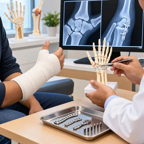
A nondisplaced, stable Rolando fracture can be managed without surgery in a thumb-spica splint or cast. Daily care means elevating the hand, using prescribed pain control, avoiding gripping and impact, attending repeat imaging, and keeping scheduled follow-up so loss of alignment is detected early.
Displacement, angulation, shortening, rotation, significant joint incongruity or instability make fixation safer. Surgery also becomes more likely after an open injury, failed cast alignment, or loss of reduction during follow-up. The choice depends on fragment size, comminution, soft-tissue condition and whether the reduction remains stable.
| Option | What it means | Main trade-off or use |
|---|---|---|
| Closed reduction with percutaneous K-wires | Realign the fracture through small skin openings and hold it with pins | Less invasive, but pins need site care and can become infected |
| Open reduction and internal fixation | Expose the joint and secure fragments with screws or a plate | Provides direct control, but requires an incision and implanted hardware |
| External fixation or ligamentotaxis | Use an outside frame and tensioned soft tissues to maintain alignment | Reserved for selected complex fractures whose small fragments cannot hold reliable screws |
The best Rolando fracture treatment is the one that restores and maintains the thumb CMC joint surface; no method suits every fracture. A cast can fail when fragments shift, while fixation carries wound, hardware, stiffness and infection risks. Regenerative injections do not restore an unstable joint surface or correct mechanical incongruity.
What thumb recovery looks like from protection to strengthening
Protection usually lasts about 4–6 weeks in a cast or splint, but your surgeon sets the timing from fracture stability, fixation, examination findings and repeat X-rays. Less pain does not mean the thumb is ready for unrestricted gripping; early overuse can shift the joint or irritate pins and hardware.
- Protect the thumb during the prescribed period. Keep heavy pinch, forceful gripping and impact loading out of daily activity. Use light tasks only when cleared, even if pain has settled.
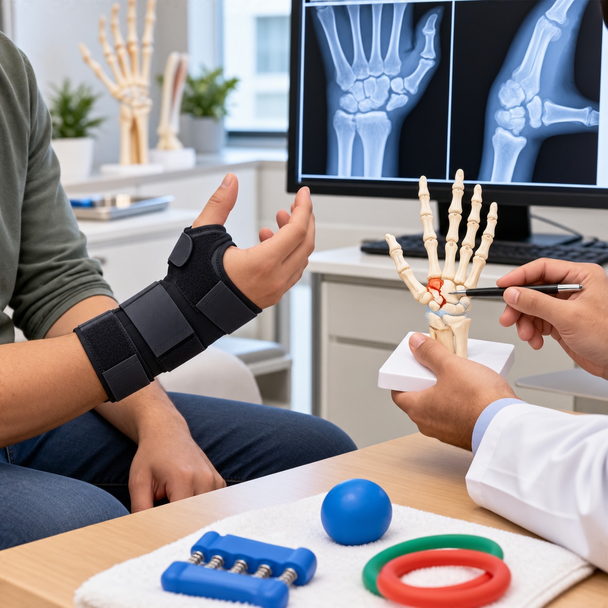
- Begin hand therapy after adequate stability is confirmed. Treatment may include thumb, wrist and finger range-of-motion exercises, swelling or scar care, opposition, pinch and grip training, followed by gradual work or sport simulation.
- Progress from light daily use to resistance, heavier gripping and impact activities in stages. Do not add aggressive self-directed stretching before clearance; it can cause loss of reduction, stiffness or irritation around fixation.
- Treat repeat X-rays as one milestone, not the finish line. Radiographic union can occur while swelling, tenderness, limited movement and reduced pinch strength remain; useful strength and confidence often improve over several months.
Rolando fracture recovery in Bengaluru follows the same principle: protect the joint first, then rebuild movement and load tolerance. Contact the treating team if pain increases after improving, the thumb becomes numb or pale, or swelling and stiffness worsen.
Possible problems include malunion, persistent CMC incongruity, stiffness, tendon or nerve irritation, pin-site infection, complex regional pain syndrome and post-traumatic CMC arthritis. Cartilage damage at the original injury can cause arthritis even after technically successful fixation.
How to choose follow-up care in Bengaluru and know when to call
Choose a service that can assess a hand injury promptly, not one that only schedules a routine orthopaedic visit. For an intra-articular thumb fracture in Bengaluru, confirm access to dedicated thumb X-rays, reduction and fixation when required, repeat X-rays, and structured hand-therapy follow-up.
Dr Harish Madrekar's Orthopaedic Clinic is one Bengaluru option for assessment and continuing orthopaedic care; ask how the team will monitor alignment and function rather than assuming one operation suits every Rolando fracture.
| Care pathway | Confirm before booking | Why it matters |
|---|---|---|
| Stable fracture in cast | Repeat X-rays and review dates | Loss of alignment can be missed as swelling falls |
| Reduced or fixed fracture | Wound or pin checks, imaging and therapy | Early problems can affect fixation and movement |
| Complex or unclear injury | CT access and orthopaedic trauma referral | Small fragments and joint alignment need better mapping |
Tell the treating team about smoking or nicotine use, diabetes, blood-thinning medication, numbness and any skin injury. These details can change fixation timing, wound care and healing advice.
Call the treating team promptly rather than waiting for the next appointment if you develop:
- Increasing pain after it had improved
- Spreading redness, drainage or fever
- New numbness
- A pale or blue thumb
- Inability to move the thumb
- A cast that becomes excessively tight
During Rolando fracture recovery in Bengaluru, early pain relief does not prove that the joint is ready for gripping or impact. Follow the scheduled imaging and therapy plan.
Related service
FRACTURE SURGERY Rolando Fractur -intra-articular fracture at the base of the first metacarpal Rolando Fracture Surgery A Rolando fracture is a complex, intra-articular fracture at the base of the first metacarpal involving the thumb... View service → |
Frequently asked questions
How does a Rolando fracture happen, and which symptoms need assessment?
A Rolando fracture can follow a fall onto an outstretched hand, axial force through the thumb, a road-traffic collision or a sports injury. Seek assessment for pain and swelling at the thumb base, bruising, deformity, reduced movement, numbness or weak pinch.
How do clinicians confirm a Rolando fracture and judge its stability?
Clinicians examine alignment, swelling, sensation, circulation and thumb movement, then use hand X-rays to identify the T- or Y-shaped intra-articular fracture. CT imaging can clarify small fragments and joint-surface displacement when X-rays do not show the full pattern.
When is a cast enough, and when is fixation safer?
A cast may be appropriate when the fracture is well aligned and remains stable during follow-up imaging. Fixation is safer when fragments are displaced, the joint surface is incongruent, the fracture is unstable or alignment cannot be maintained in a cast.
What does thumb recovery look like after a Rolando fracture?
Recovery progresses from protection in a cast or splint to guided thumb and wrist movement, followed by grip, pinch and strengthening exercises after healing permits. Your clinician sets activity limits and checks progress with examination and imaging.
When should you call your follow-up team during recovery?
Call promptly for increasing pain or swelling, a tight or damaged cast, numbness, blue or pale fingers, fever, wound drainage, new deformity or loss of thumb movement. Attend scheduled reviews so alignment and healing can be checked.
Keywords